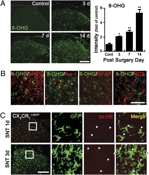
Fig. 1.
ROS are produced in the spinal cord microglia after L5 SNT. (A) Spinal cord sections of uninjured and SNT-injured mice at 3, 7, and 14 d (each group, n = 4) after L5 SNT were used for 8-OHG immunostaining. (Scale bar, 200 μm.) The 8-OHG+ cells were detected in spinal cord dorsal horn only after SNT. The intensity of 8-OHG immunoreactivity was measured, and the mean ± SEM values are presented (*P < 0.05; **P < 0.01). (B) 8-OHG+ (green) cells were immunostained with cellular markers (red) for neurons (MAP2), microglia (Iba-1), astrocyte (GFAP), and oligodendrocyte precursor cells (NG2). (Scale bar, 50 μm.) (C) Superoxide generation in SNT-injured CX3CR1+/GFP mice was detected with oxidized-hydroethidine (ox-HE) staining. Ox-HE signals (red) were mainly detected in the GFP+ microglia (green) and in the ipsilateral spinal cord of 1 and 3 d post-SNT. Enlarged images are shown in rectangles; ox-HE+ cells are marked with white triangles. (Scale bar, 200 μm.)

