Fig. 2.
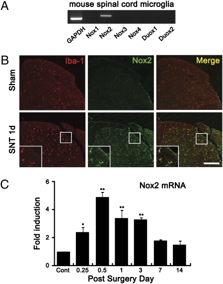
Nox2 expression is increased in spinal cord microglia after SNT. (A) Transcripts of Nox1, Nox2, Nox3, Nox4, Duox1, and Duox2 in primary mouse spinal cord microglia were detected by RT-PCR. (B) Spinal cord sections were immunostained with Iba-1 and Nox2 antibodies. Nox2 immunoreactivity signals were increased in Iba-1+ microglia in the spinal cord dorsal horn at 1 d after SNT. (Scale bar, 200 μm.) (C) Nox2 mRNA expression in mouse spinal cord after L5 SNT was measured by real-time RT-PCR. Total RNA was isolated from L5 spinal cord tissues of uninjured mice (n = 3) and SNT-injured mice at 6 h, 12 h, and 1, 3, 7, and 14 d after surgery (each group, n = 3). The Nox2 transcript level at each time point was normalized to the level of GAPDH and is presented as fold induction, compared with the Nox2 level of uninjured mice. Data are expressed as mean ± SEM (*P < 0.05; **P < 0.01).

