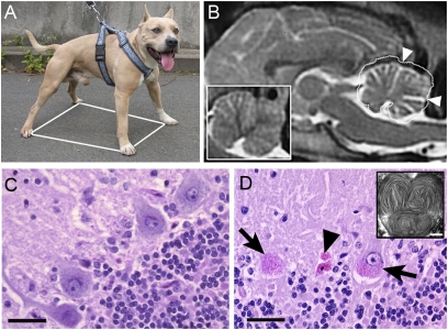
Fig. 1.
Clinical and histopathological features of the disease. (A) The wide-based stance (white polygon) of a 5-y-old affected AST illustrates loss of motor coordination. (B) Sagittal 2-T weighted MRI of the brain from a 6-y-old AST through the cerebellum. (Inset) A similar image at the same scale of the cerebellum from an age-matched healthy AST, the outline of which has been projected on the cerebellum of the main image (white dotted line). A reduction of gray matter is demonstrated by the enlarged sulci (arrowheads). (C and D) Transverse sections of the cerebellum from healthy dogs (C) and affected dogs (D), stained with PAS reagent and counterstained in Mayer's hematoxylin solution. (C) In a normal cerebellum, the large PAS-negative Purkinje neurons lie between the molecular (top left) and granular (bottom right) layers. (D) In the cerebellum from an affected dog, massive Purkinje cell loss results in a blurry line. The remaining Purkinje neurons (arrows) or neuron remnants (arrowheads) accumulate perinuclear PAS-positive granular material. (Inset) Affected Purkinje neurons imaged on transmission electron microscopy show accumulated lysosomal material composed of concentric straight or curved profiles with alternating clear and dense bands. (Scale bars: 50 μm for the sections; 250 nm for the inset.)

