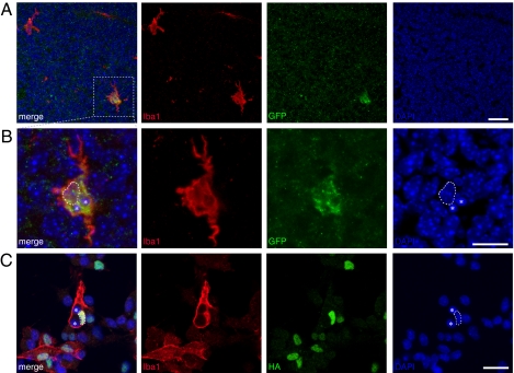
Fig. 5.
PU.1 reprogrammed neural stem cell–derived cells display monocyte traits. (A and B) Neural stem cell–derived CD45+/GFP+ lenti-PU.1-GFP–transduced cells were transplanted into the lateral ventricle in E15 mouse embryos. A GFP-labeled cell (shown in the box in A and in higher magnification in B) expresses the monocyte marker Iba1 and displays a morphology indistinguishable from that of a resident microglial cell at 4 d after transplantation. The GFP-labeled cell contains two vacuoles with DAPI+-condensed chromatin (asterisks), indicating phagocytosis of cellular debris. The dashed lined delineates the cell's own nucleus. (C) Iba1-expressing PU.1-transduced neurosphere cells in vitro were found to have phagacytotic capacity and to contain condensed DAPI+ cellular debris (asterisks). The dashed lined delineates the cell's own nucleus. (Scale bar: 25 μm.)

