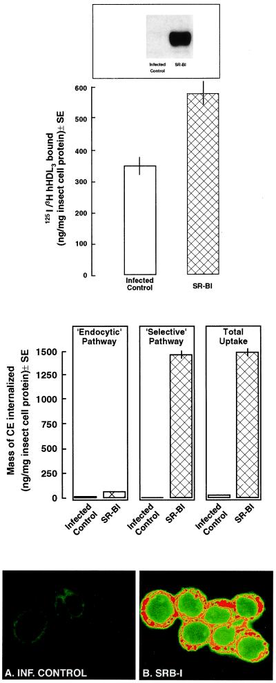
Figure 3.
Binding of 125I-/[3H]hHDL3 and selective CE uptake in Sf9 cells expressing SR-BI. (Top) Sf9 cells infected with recombinant baculovirus coding for SR-BI or infected control incubated for 5 h with 125I-DLT-[3H]COE-hHDL3 plus unlabeled hHDL3. Specific binding was defined as the difference between total and nonspecific binding (in the presence of 500 μg/ml unlabeled hHDL3). The expression of SR-BI protein was confirmed by immunoblotting (see blot at the top of the figure in which 20 μg of cellular lysate was loaded). (Middle) The mass of CEs internalized via the endocytic and selective pathways is shown for infected control and SR-BI-expressing cells. Determination of the mass of CEs internalized via the endocytic and selective pathways is described in Materials and Methods. (Bottom) Infected control and SR-BI-expressing cells were incubated with reconstituted BODIPY-CE-HDL to microscopically identify CE uptake. By using confocal microscopy, the infected control cells (A) show no fluorescence, whereas SR-BI-expressing cells (B) show patchy areas of high (red) or medium (orange) uptake of CEs.

