Fig. 4.
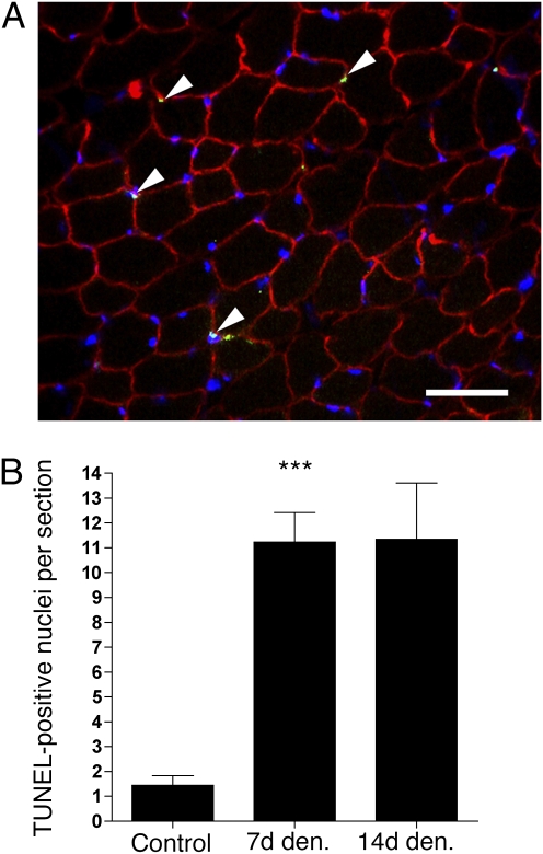
Apoptosis in hypertrophied muscle after denervation (Den.). (A) Triple staining with Hoechst 33342 (blue), TUNEL (green), and antibodies against dystrophin (red). Note that all apoptotic nuclei (arrows) are outside the muscle cells; hence, they are either satellite or stroma cells. (Scale bar: 50 μm.) (B) Number of TUNEL-positive nuclei per section. Each column point represents the mean ± SEM (n = 5–8 sections from four muscles). ***Statistical significance (P < 0.0001).

