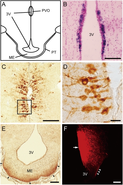
Fig. 1.
Localization of Opsin 5 in the PVO and its projections to the external zone of the median eminence. (A) Schematic drawing of the quail mediobasal hypothalamus. (B) In situ hybridization of OPN5 mRNA in the PVO. (C–E) Representative image of Opsin 5-like immunoreactivity in PVO neurons that contact the CSF (C and D) and fibers in the external zone of median eminence (ME) (arrowhead) adjacent to the pars tuberalis (PT) of the pituitary gland (E). Note that the angles of the sections are slightly different between B and C (Fig. S7). (F) The neural tracer DiI was applied to the PVO (arrow) and labeled fibers in the external zone of the ME (arrowheads). (Scale bars: 10 μm, D; 100 μm, B, C, and E; and 200 μm, F.) 3V, third ventricle.

