Fig. 4.
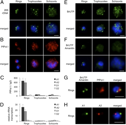
Disruption of rDNA nucleolar clustering and decrease of rDNA transcription in replicative and mitotic stages. (A) FISH analysis of 28S rDNA (green) during 48-h blood-stage cycle. In ring stage (0–18 h postinvasion), rDNA is clustered at one pole of the nucleus, whereas in replicative and dividing stages (trophozoite, 18–38 h and schizont, 38–48 h), the individual rDNA units appear dispersed at multiple sites. (B) Immunofluorescence analysis of PfPol I (red). Ring-stage parasites show a half-moon perinuclear nucleolus. In trophozoites, PfPol I shows an additional diffuse pattern in the nucleoplasm and cytoplasm. In schizont segmented forms, PfPol I appears to relocalize at the nuclear periphery (Fig. S4). (C) ChIP analysis of PfPol I occupation on the individual rRNA genes (A1, A2, S1, and S2) during blood-stage cycle (mean ± SEM; n = 4). (D) Relative rRNA gene transcription throughout the blood-stage cycle. S2 values were set to 1 (mean ± SEM; n = 3). (E) Br-UTP labeling of nascent RNA (green, mean ± SD: rings 5.51 ± 1.57 foci/nucleus, n = 69; trophozoites 11.09 ± 3.30, n = 56; schizont-stages >20 foci). (F) Br-UTP labeling of nascent RNA in the presence of α-amanitin (green; rings 2.02 ± 0.69 foci/nucleus, n = 61). (G) Colocalization of α-amanitin–resistant foci (green) with nucleolus (red, PfPol I). (H) Two-color DNA FISH of A1 (green) and A2 (red) rRNA genes in ring-stage parasites (colocalization 23.30%, n = 103). In all images, parasite nuclei are shown by DAPI staining (blue). (Scale bars, 2 μm.)

