
Figure 4.
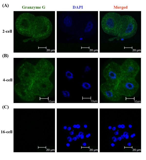
Immunoflourescence staining of Granzyme G protein expression and subcellular localization at different stage embryos. (A) Two-cell stage mouse embryos (n = 6). (B) Four-cell stage mouse embryos (n = 6). (C) Sixteen-cell stage mouse embryos (n = 5). The left panels show embryos stained with a granzyme G-specific primary antibody and FITC-conjugated secondary IgG antibody. The middle panels show embryos stained with DAPI for nuclear localization. The right panels show merged images of grazyme G and DAPI staining.