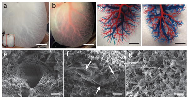
Figure 2.
DLM retains intact lobular structure and vascular bed. (a) Representative photograph of decellularized left lateral and median lobes of rat liver, with the vascular tree visible. (b) The vascular tree, after perfusion with Allura Red AC dye. (c,d) Corrosion cast model of left lobe of a normal liver (c) and the DLM (d), with portal (red) and venous (blue) vasculature. (e–g) SEM images of a vessel (e), a section featuring bile duct–like small vessels (arrows) (f), extracellular matrix within the parenchyma (g), with hepatocyte-size free spaces. Scale bars: 10 mm (a,b), 5 mm (c,d) and 20 μm (e–g).

