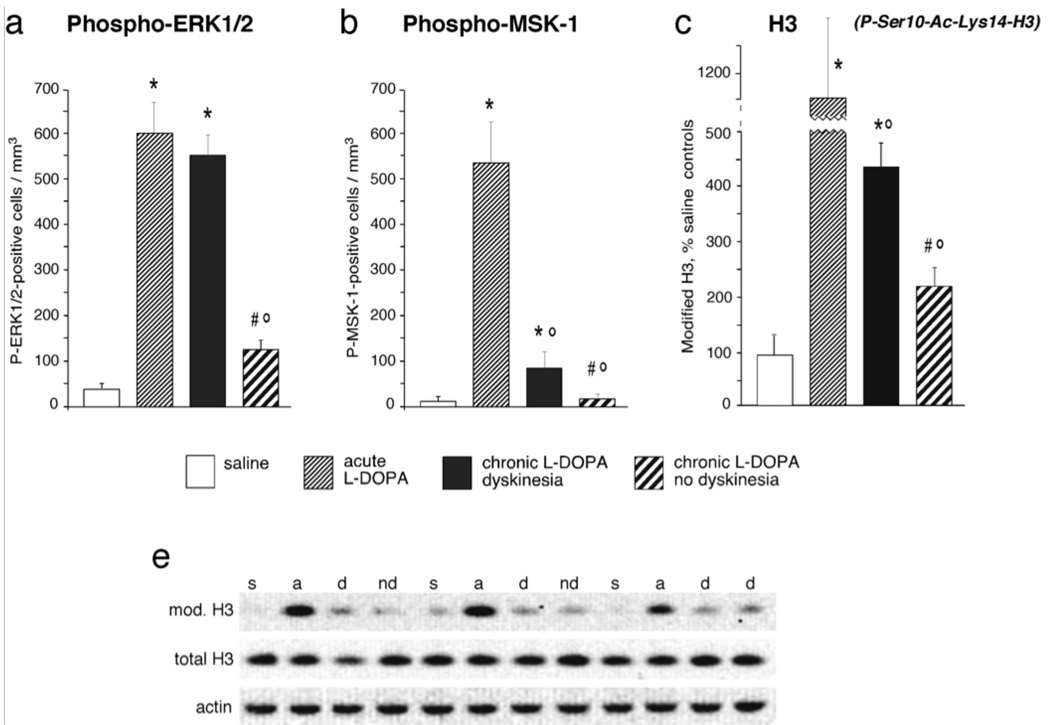
Figure 2. Deficient densensitization of DA receptor-dependent signaling in the dyskinetic striatum.
Acute treatment with l-DOPA results in a supersensitive activation of cAMP- and MAPK-dependent pathways signaling to the nucleus, here exemplified by the phosphorylation of ERK1/2 (on Thr202-Tyr204), MSK-1 (on Ser376) and by a modification of histone 3 that has been linked to chromatin remodelling during active gene transcription (phospho[Ser10]-Acetyl[Lys14]-H3). Chronic l-DOPA administration normalizes these supersensitive responses only in animals that remain free from dyskinesia during the treatment (non-dyskinetic group). Levels of signaling pathway activation remain significantly elevated above control values in dyskinetic animals. Thus, a shift towards normal molecular responses during chronic l-DOPA treatment is associated with a resistance to dyskinesia, while persistent supersensitivity is a correlate of LID. These data show both published and unpublished results obtained from 6-OHDA-lesioned rats, which received chronic (10–12 days) or acute treatment with l-DOPA (l-DOPA methyl ester, 10 mg/kg/dose combined with 15 mg/kg/dose of benserazide), or physiological saline, and were killed 30 minutes post injection. Data in (a) and (b) represent counts of immunoreactive neurons in the lateral part of the DA-denervated caudate–putamen, from animals reported in Westin et al. (2007), and five chronically l-DOPA-treated non-dyskinetic rats from the same study (not reported in the paper). Data in (c) show results from Western immunoblotting analysis. The optical density on specific immunoreactive bands was normalized to the corresponding β-actin bands, and results from each group were expressed as a percentage of saline control values. A Western blot showing bands immunoreactive for phospho[Ser10]-Acetyl[Lys14]-histone 3, total histone 3 and beta-actin is shown in (d). Full descriptions of the experimental procedures can be found in Westin et al. (2007) (for a, b) and Schroeder et al. (2008) (for c, d). Abbreviations in (d): s, saline control; a, acute l-DOPA; d, chronic l-DOPA/dyskinesia; nd, chronic l-DOPA/no dyskinesia). Values indicate group means ± SEM from 3 to 10 animals per group, which were statistically compared using one-factor analysis of variance and post-hoc Newman– Keuls test. P < 0.05 vs. *, saline-treated 6-OHDA-lesioned controls; °, acute l-DOPA; #, chronic l-DOPA with dyskinesia.

