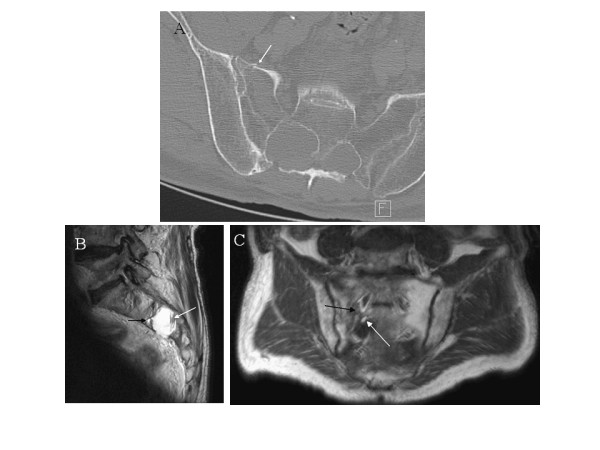
Figure 2.

Sacral cyst CT-scan and MRI image. A. Axial sacral CT-scan: left sacral fracture extending to the S2 radicular cyst (presence of a contralateral cyst at the same level). B. Sagittal T2-weighted sacral MR image: S2 radicular cyst with two liquids: cerebrospinal fluid with blood sediments (white arrow) and fat droplet (black arrow). C. Coronal T1-weighted sacral MR image: left sacral fracture extending to the radicular cyst (black arrow) which contains cerebrospinal fluid and fat droplets at the bottom (white arrow).
