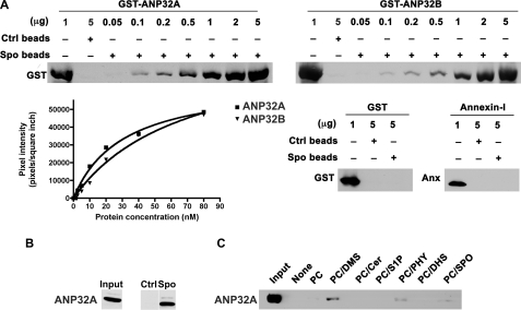
FIGURE 2.
Interaction of ANP32A and ANP32B with sphingolipids. A, different amounts of purified GST-ANP32B, GST-ANP32A, GST, or annexin I were incubated with sphingosine or control beads (1 mg) for 10 min, eluted with SDS sample buffer, and separated by SDS-PAGE. Western blot analysis was performed using an α-GST antibody or α-annexin I antibody. Band intensities were quantitated using a densitometer. B, HEK293T cell extract (1 mg) was incubated with sphingosine beads or control beads (1 mg), washed, eluted with SDS sample buffer, and separated by SDS-PAGE. Western blot analysis was performed using an α-ANP32A antibody. Input represents 50 μg of whole cell lysate. C, GST-ANP32A bound to indicated liposomes (250 μm) was centrifuged, and ANP32A present in the pellet was detected by immunoblot analysis.

