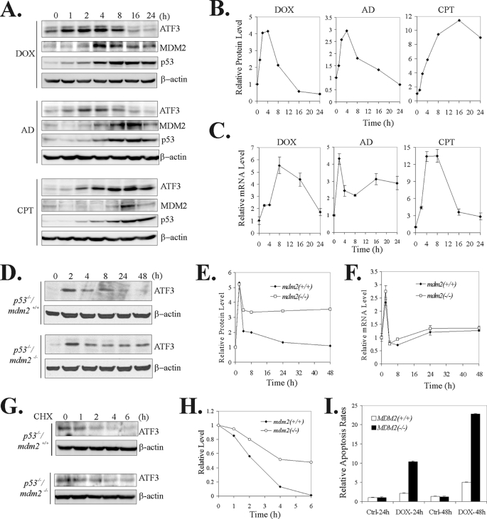
FIGURE 1.
MDM2 regulates ATF3 levels in the DNA damage response. A and B, A549 cells were treated with 0.4 μg/ml DOX, 2 nm AD, or 1.5 μm CPT and lysed at the indicated times for immunoblotting (A). Densitometry was used to quantitate ATF3 levels (B). C, A549 cells were treated as in A. Total RNA was prepared and subjected to real-time RT-PCR to quantify ATF3 mRNA levels. D–F, mdm2-wild-type (p53−/−/mdm2+/+) or -deficient (p53−/−/mdm2−/−) MEF cells were treated with 0.4 μg/ml DOX for the indicated times and then subjected to immunoblotting (D) or real-time RT-PCR (F). ATF3 levels were quantitated by densitometry, and the results are shown in E. G and H, indicated cells were treated with 0.4 μg/ml DOX for 8 h. After removal of DOX, 100 μg/ml cycloheximide (CHX) was added into culture medium. Cells were harvested at the indicated times for immunoblotting. ATF3 levels were quantitated by densitometry, and the results are shown in H. I, p53−/−/mdm2+/+ or p53−/−/mdm2−/− MEF cells were treated with 0.4 μg/ml DOX for 24 and 48 h and stained with propidium iodide for flow cytometry analysis as described previously (3). Percentages of subG0/G1 cells were used to calculate the folds of increase in apoptosis rates after DOX treatments.

