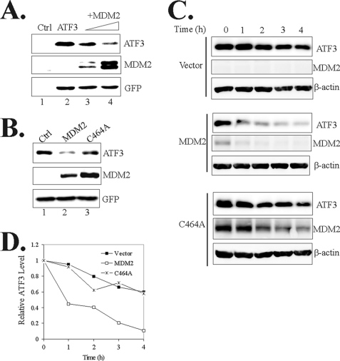
FIGURE 3.
MDM2 promotes ATF3 degradation. A, H1299 cells were transfected with ATF3, GFP, and increasing amounts of MDM2 as indicated. Cells were lysed, and GFP expression levels were quantitated using a fluorescence spectrophotometer to normalize transfection efficiencies. Normalized cell lysates were then subjected to immunoblotting. B, H1299 cells were transfected as indicated and subjected to immunoblotting as in A. C and D, H1299 cells were transfected and treated with 100 μg/ml cycloheximide for different time. ATF3 levels were quantitated by densitometry, and the results are shown in D.

