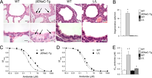
FIGURE 6.
Regulation of ENaC in airway epithelia is abnormal in βENaC-Tg mice but preserved in Liddle mice. A, airway histology from neonatal (3-day-old) βENaC-Tg mice, L/L mice, and their respective WT littermates. Sections were stained with H&E and evaluated for degenerative airway epithelial cells (arrows). Scale bars, 20 μm (upper panels) and 10 μm (lower panels). B, summary of airway epithelial necrosis as determined from the number of degenerative epithelial cells per mm of the basement membrane. n = 3–5 mice for each group. *, p ≤ 0.01 compared with WT. C–E, amiloride dose-response curves (C and D) and summary of IC50 values (E) obtained from tracheal tissues of βENaC-Tg mice, L/L mice, and respective WT littermates. n = 6–12 mice/group. *, p < 0.01 compared with WT mice; †, p = 0.001 compared with L/L mice. Error bars, S.E.

