FIGURE 1.
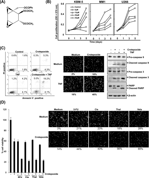
Crotepoxide inhibits the proliferation of leukemic cells and potentiated the apoptotic effects of TNF and chemotherapeutic agents. A, shown is the chemical structure of crotepoxide. Ph, phenyl. B, crotepoxide inhibited the proliferation of KBM-5, MM1, and U266 cells. Cells were seeded in 96-well plates and treated with the indicated concentrations of crotepoxide. Cell proliferation was analyzed by MTT assay on days 1, 3, and 5. C, crotepoxide enhanced TNF-induced apoptosis. KBM-5 cells were pretreated with crotepoxide (50 μm) for 2 h then treated with TNF (1 nm) for 24 h. Cell death was determined by fluorescence-activated cell sorting using annexin V/propidium iodide staining (left panel) and by live/dead assay (middle panel). Cleavage of caspase-9 and −3, and poly(ADP-ribose) polymerase was determined by Western blotting in whole-cell extracts of crotepoxide- and TNF-treated cells (right panel). D, crotepoxide potentiated cytotoxicity induced by 5-flurouracil (5-FU), cisplatin (Cis), thalidomide (Thal), and velacade (Vel) is shown. Five thousand cells were seeded in triplicate in 96-well plates, pretreated with crotepoxide (50 μm) for 2 h, and then incubated with chemotherapeutic agents for 24 h. Cell viability was then analyzed by MTT assay (left panel). Crotepoxide also potentiated chemotherapy-induced apoptosis. KBM-5 cells (1 × 10⁶) were pretreated with crotepoxide (50 μm) for 2 h then treated with TNF (1 nm) for 24 h. Cell death was analyzed by a live/dead assay (right panel).

