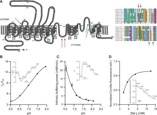
FIGURE 1.
A, proposed topological model for the mouse slc4a10-derived polypeptide based on the similar model from slc4a1, Ae1. The mutated amino acids are highlighted in red and by arrows. Right panels show the amino acid alignment of the hinge-like loops of slc4a-derived polypeptides. Mutated amino acids are marked with arrows. (Modified with permission from J. Casey.) B, calibration of excitation fluorescence ratio (I495/I440) into pHi values. Inset shows fluorescence ratio trace and extracellular pH values from one experiment. C, determination of pHi buffering capacity, βint. Inset shows pHi trace and extracellular [NH4+ + NH3] values from one experiment. D, calibration of intracellular CoroNa fluorescence to [Na+]i values. Inset shows fluorescence recording and extracellular [Na+] values from one experiment. Error bars = S.E.

