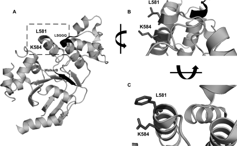
FIGURE 3.
A, stereo ribbon diagram of human F508del-NBD1 structure (Protein Data Bank code 2BBT) revealing the positions of Leu581 and Lys584 (highlighted by the square). The LSGGQ motif and the Walker B motif are also shown (black ribbon and arrow, respectively). B, close-up view of the same structure as in A (human NBD1) in the vicinity of Lys584 (dark gray) showing the interaction of its side chain with that of Leu581 (dark gray). C, similar close-up view but in the mouse NBD1 structure (Protein Data Bank code 1RXO) showing the interaction between the side chains of the corresponding mouse residues, namely Glu584 and Phe581 (both in dark gray). In B and C, the side chains of residues 581 and 584 are shown (dark gray).

