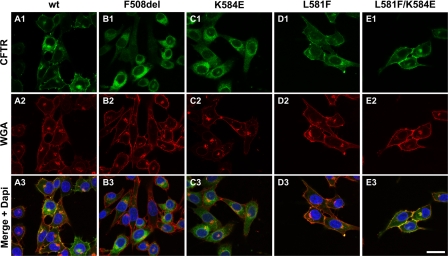
FIGURE 5.
Immunolocalization of CFTR constructs in BHK cells. BHK cells expressing the indicated CFTR variants grown at 37 °C were analyzed by immunofluorescence using the anti-CFTR antibody 570 (green; upper panels) and the anti-wheat germ agglutinin (WGA) coupled with Texas Red (red; middle panels). The lower panels show the merged images with nuclei stained blue with DAPI. Data shown are representative of n = 4 experiments. Bar, 25 μm.

