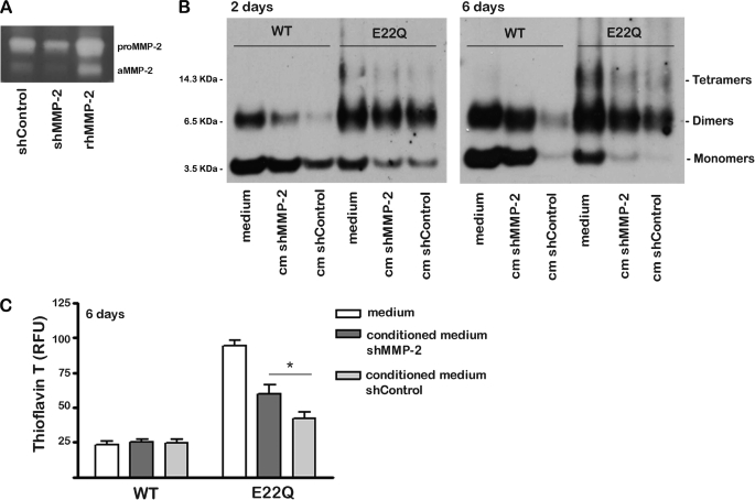
FIGURE 7.
In vitro digestion of Aβ peptides by conditioned media of shMMP-2 and shControl cells. Aβ peptides (WT and E22Q) were incubated in vitro for 2 and 6 days at 37 °C with conditioned media from amyloid-unchallenged shMMP-2 and shControl cells. Aβ degradation was evaluated by Western blot analysis and thioflavin-T binding, as described under “Experimental Procedures.” As controls, the respective Aβ peptides were incubated under identical conditions with culture media that had not been preincubated with the corresponding cell lines. A, gel zymography of conditioned media from amyloid unchallenged shMMP-2 and shControl cells grown to confluence to maximize endogenous MMP-2 secretion levels is shown. The electrophoretic bands corresponding to rhMMP-2, both the pro-form and the activated enzyme, are shown for comparison. aMMP-2, active MMP-2. B, shown is Western blot analysis of Aβ peptides after incubation with the respective conditioned media, probed with a combination of 4G8 and 6E10 anti-Aβ. Lanes labeled medium represent media that had not been in contact with the respective cell lines; cm indicates conditioned medium. Electrophoretic mobility of the molecular weight standards is indicated on the left of the panel. C, shown is a thioflavin-T fluorescence assay of the respective conditioned media. RFU, relative fluorescence units. (Bars represent the mean ± S.E. of three independent experiments. * indicates statistically significant differences with p < 0.05.

