FIGURE 8.
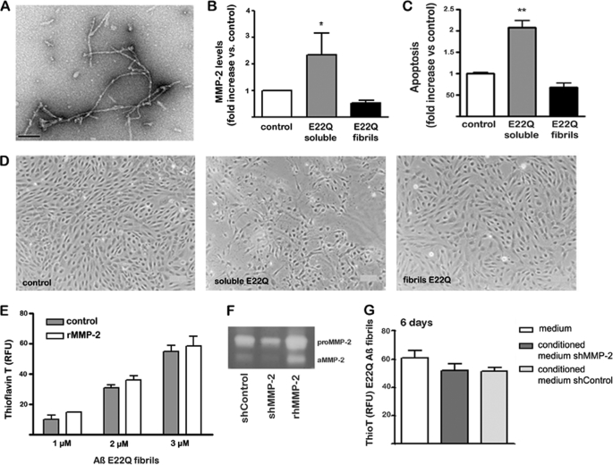
AβE22Q fibrillar assemblies fail to elicit MMP-2 release and activation or an apoptotic response in microvascular endothelial cell cultures. EC cultures were challenged with AβE22Q soluble and fibril-enriched assemblies for 3 days followed by evaluation of MMP-2 secretion, assessment of Aβ fibrillar components in culture supernatants by thioflavin-T-binding assays, and estimation of apoptosis induction by morphological studies and Cell Death ELISA. A, electron microscopy images illustrating AβE22Q fibrillar assemblies were prepared as described under “Experimental Procedures”; the bar represents 100 nm. Note: please refer to Fig. 1A for EM visualization of AβE22Q-soluble assemblies. B, evaluation of MMP-2 secretion by SearchLight® human MMP antibody arrays is shown. Results are expressed as -fold increase with respect to control cells incubated in the absence of Aβ peptides. Bars represent mean ± S.E. of triplicate experiments; * indicates statistically significance differences with p < 0.05. C, evaluation of apoptosis induction by nucleosome quantitation via Cell Death ELISA is shown. Apoptosis rate is represented in -fold change with respect to untreated control cells. Bars indicate the mean ± S.E. of three independent experiments; ** highlights statistically significant differences with p < 0.01. D, contrast phase microscopy images at 100× magnification exemplify changes in cell density and morphology after Aβ treatment. E, shown is a thioflavin-T fluorescence assay of AβE22Q fibrils after 24 h in vitro incubation with rhMMP-2; as controls, the fibrillar assemblies were incubated under identical conditions in the absence of the enzyme. Bars represent the mean ± S.E. of at least three independent experiments. F, gel zymography of conditioned media from untreated shMMP-2 and shControl cells is shown; the electrophoretic bands corresponding to rhMMP-2, both the pro-form and the activated enzyme, are also shown as control. aMMP-2, active MMP-2. G, shown is a thioflavin-T fluorescence assay of AβE22Q fibrils after in vitro incubation for 6 days with conditioned media from shMMP-2 and shControl cells. As negative controls, AβE22Q fibrils were incubated with plain media that had not been in contact with cells. Results are expressed as the mean values ± S.E. of at least three independent experiments. RFU, relative fluorescence units.

