FIGURE 8.
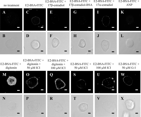
Specific binding of 17β-estradiol-BSA-FITC at the hepatocyte plasma membrane. Isolated hepatocytes were seeded onto coverslips and allowed to attach for 24 h. Cells were treated with either no treatment (A and C), 50 μm 17β-estradiol (E), 50 μm 17β-estradiol-BSA (G), 50 μm 17α-estradiol (I), 20 μm ANP (K), 100 μm digitonin (M), 100 μm digitonin plus 50 μm ICI 182,780 (ICI) (O), 100 μm digitonin plus 100 μm ICI (Q), 50 μm ICI 182,780 (S), 100 μm ICI 182,780 (U), or 50 μm G-1 (W) for 20 min at 4 °C. 1 μm 17β-estradiol-BSA-FITC (E2-BSA-FITC) was then added (C–W), and the cells were incubated for a further 20 min at 4 °C. Cells were then fixed and mounted onto coverslips. Corresponding phase images (B–X) demonstrate general cell morphology. The scale bar represents 10 μm, and data are representative of several experiments from at least 3 independent hepatocyte preparations.

