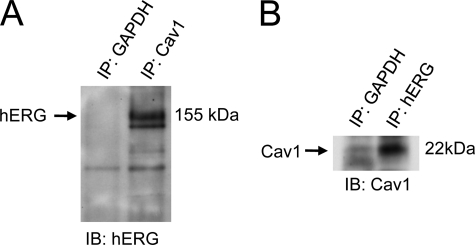
FIGURE 3.
Co-immunoprecipitation of hERG and Cav1. hERG-HEK cells were lysed, and samples of 0.5 mg of protein were used for immunoprecipitation (IP) using an anti-Cav1 antibody (A) or an anti-hERG antibody (B). The immunoprecipitated protein was then immunoblotted (IB) using an anti-hERG antibody (A) or anti-Cav1 antibody (B). An anti-GAPDH antibody was used as a negative control to avoid nonspecific binding between target protein and beads.

