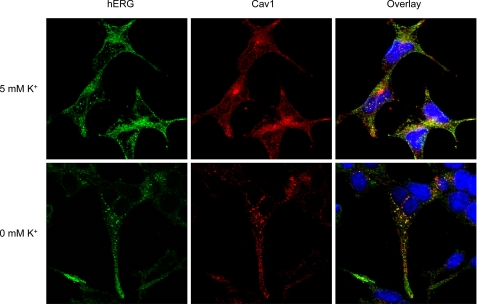
FIGURE 4.
Colocalization between hERG and Cav1 in hERG-HEK cells during endocytosis. hERG-HEK cells exposed to 5 or 0 mm K+ MEM for 3 h were fixed and permeabilized. The cells were treated with goat anti-hERG primary and Alexa Fluor 488-conjugated anti-goat secondary antibodies to detect hERG (green) and with rabbit anti-Cav1 primary and Alexa Fluor 546-conjugated anti-rabbit secondary antibodies to detect Cav1 (red). Nuclei were stained using Hoechst 33342.

