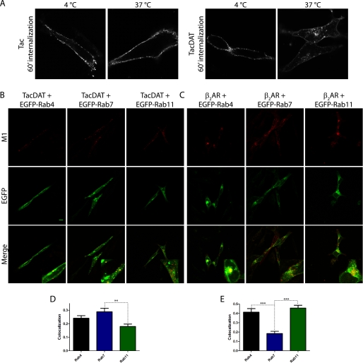
FIGURE 4.
TacDAT is constitutively internalized in the dopaminergic cell line 1Rb3An27 and co-localizes primarily with EGFP-Rab7 and intermediately with EGFP-Rab4. A, experiment as in Fig. 2A on 1Rb3An27 cells expressing Tac or TacDAT. Pictures are representative of several experiments. B, fluorescence co-localization between TacDAT and the EGFP-tagged endosomal marker Rab4, Rab7, or Rab11 after 1 h of internalization in the presence of M1 antibody. C, fluorescence co-localization of FLAG-tagged β2-adrenergic receptor (β2AR), a bona fide recycling membrane protein, with EGFP-Rab4, -Rab7, or -Rab11 after 1 h of isoproterenol (10 μm)-induced internalization. In both B and C, upper panels show Alexa Fluor 568 signal (M1), middle panels EGFP signal, and lower panels show the overlay of the two channels. D and E, quantification of fluorescence co-localization in B and C between internalized TacDAT (D) or internalized FLAG-tagged β2-adrenergic receptor (E) and the EGFP-tagged endosomal marker Rab4, Rab7, or Rab11 (means ± S.E., p < 0.01, one-way ANOVA, Bonferroni's multiple comparison test). Co-localization data were analyzed from 30 images of each condition. Representative images are shown in B and C.

