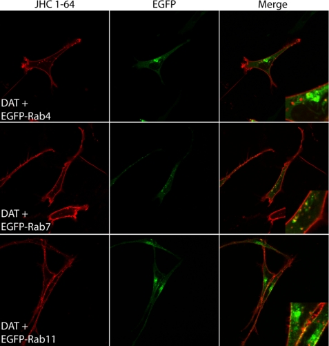
FIGURE 5.
Visualization of constitutive DAT internalization in live 1Rb3An27 cells using fluorescent cocaine analogue JHC 1-64 reveals predominant co-localization with EGFP-Rab7. 1Rb3An27 cells expressing DAT together with EGFP-Rab4, -Rab7, or -Rab11 were incubated with JHC 1-64 (5 nm) at 4 °C to label surface DAT and subsequently incubated at 37 °C to drive internalization of DAT. After internalization, the live cells were imaged using confocal microscopy. Left panels show rhodamine signal (JHC 1-64), middle panels show EGFP signal, and right panels show the overlay of the two channels. Images are representative of three independent experiments.

