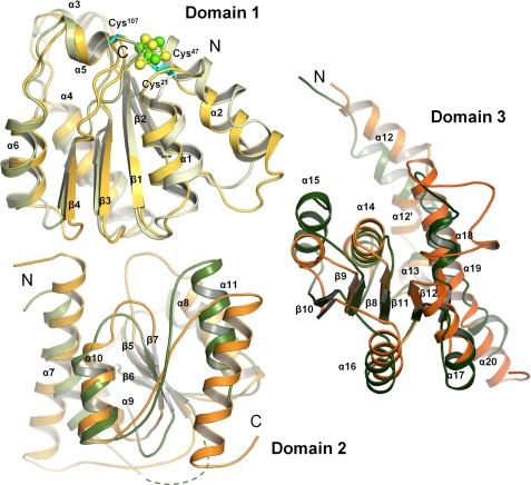
FIGURE 4.
Comparison of subdomains of ChlN and ChlB. ChlN (shades of green) and ChlB (yellow to orange) each consist of three similar subdomains. Each subunit bears a central, parallel β-sheet surrounded by α-helices. The first subdomain of ChlN and ChlB serves to coordinate the [4Fe-4S] and is involved in ChlL2 binding. The second subdomain appears to have a largely structural role in positioning the remaining two domains but may also be involved in substrate recognition. The third subdomain of ChlN and ChlB is involved in forming the active site channel and in substrate recognition.

