
Figure 1.
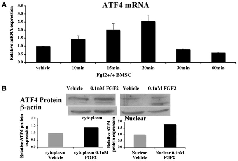
FGF2 stimulates ATF4 expression in Fgf2+/+ BMSCs. Freshly isolated BMSCs were cultured in αMEM 10% FBS for 6 ds, serum deprived 14 hrs, then (A) treated with 0.1 nM FGF2 or vehicle at indicated times, followed by RNA extraction. ATF4 mRNA was quantified by quantitative real time PCR, normalized to GAPDH. (B) BMSCs cultured as above were treated with 0.1 nM FGF2 or vehicle for 3 hrs, followed by extraction of cytosolic and nuclear protein fraction and Western Blots. Western blot Bands were analyzed by NIH Image. Two or three independent experiments were conducted with similar results, and results of representative experiments are shown.