
Fig. 1.
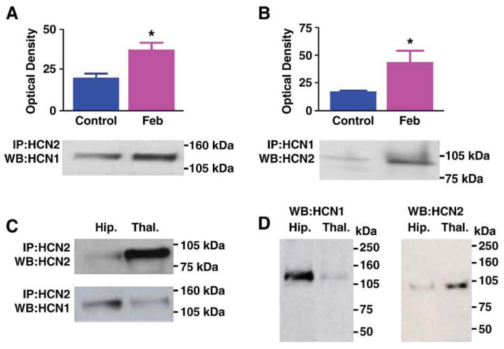
(A) Immunoprecipitation (IP) with antiserum to HCN2 followed by Western blot (WB) analysis for HCN1 shows enhanced co-association of HCN1/HCN2 in hippocampi from animals subjected to experimental prolonged febrile seizures on postnatal day 10 (Feb). The immunoreactive HCN1 band has an apparent molecular weight of approximately 110 kDa. Quantitative analysis of optical density of HCN1 immunoreactive bands reveals a significant increase in HCN1/HCN2 heteromerization 1 week after the induction of the seizures. (B) IP with antiserum to HCN1 followed by Western blot analysis for HCN2 corroborates the greater co-association of the two isoforms in the Feb group. (C) IP with antiserum to HCN2 followed by Western blot analysis for the same isoform shows greater HCN2 precipitation and immunoreactivity in thalamus compared with hippocampus (top blots). By contrast, hippocampal homogenates contain higher levels of HCN1 that is co-immunoprecipitated with the HCN2 antiserum (bottom blots). Note also different molecular weights of the HCN1 and HCN2 bands. (D) Western blots (without prior IP) for HCN1 (left) and HCN2 (right) demonstrate that the antisera used here detect HCN1 (abundant in hippocampus) and HCN2 (abundant in thalamus), and bind to antigens of the expected molecular weights, further supporting the specificity of these antisera.