Fig. 3.

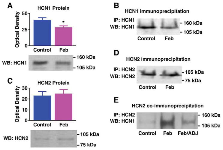
Differential regulation of the HCN1 and HCN2 protein levels promotes heteromerization: (A) Western blot (WB) showing a significant decrease on HCN1 protein levels 7 days after experimental ‘febrile’ seizures (Feb, P = 0.03). (B) Reduced HCN1 protein after seizures is apparent also when IP is carried out with anti-HCN1 followed by WB probing with the same antiserum: a fainter immunoreactive band is seen in a Feb hippocampal homogenate, compared with a hippocampus from a control animal. Note that the samples shown here are from the same hippocampi used in Fig. 1B. Thus, increased HCN1/HCN2 (Fig. 1B) is found despite reduced total HCN1 immunoreactivity. (C) HCN2 protein levels are not changed after experimental febrile seizures, as shown in the WB using anti-HCN2. (D) In accord with the WB, IP for HCN2 followed by probing for the same isoform does not lead to increased HCN2-immunoreactive band intensity. For each gel, every lane was loaded with equal amounts (25 μg) of extract from a single hippocampus. (E) When equal amounts of HCN2 protein derived from a Feb or a control hippocampus are immunoprecipitated with anti-HCN2 and then probed for HCN1, more HCN1 is co-associated with HCN2 in the Feb group. A Feb homogenate in which a mild increase of HCN2 protein occurred was subjected to WB for HCN2, compared with a control. Amount of this Feb homogenate, adjusted to include the same HCN2 quantity as in the control (Feb/ADJ) was immunoprecipitated with anti-HCN2, together with the control and the non-adjusted Feb homogenate (Feb). As shown, in Feb hippocampus, higher levels of HCN1 still co-precipitated with HCN2 even when HCN2 amounts were carefully adjusted to equal those in the control, suggesting that reduced HCN1 in the homogenate or other seizure-evoked factors govern enhanced HCN1/HCN2 co-association in the Feb group.
