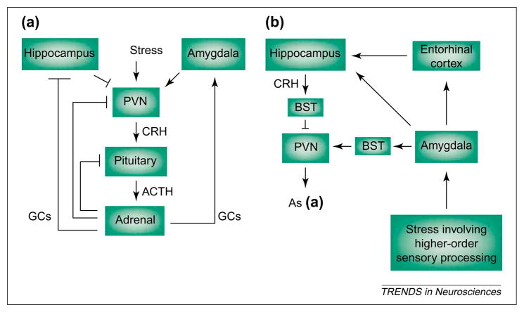
Fig. 1.
Stress-activated pathways include the neuroendocrine hypothalamic–pituitary–adrenal axis (a) and the central, limbic stress-loop (b). (a) ‘Physiological’ stress signals reach the hypothalamus, causing secretion of corticotropin-releasing hormone (CRH) from neurons of the paraventricular nucleus (PVN). CRH induces release of adrenocorticotropic hormone (ACTH) from the pituitary, and ACTH elicits secretion of glucocorticoids (GCs) from the adrenal gland. GCs cross the blood–brain barrier and activate specific receptors in hippocampus (and other CNS regions) to ‘shut off’ the neuroendocrine stress response. By contrast, GCs increase CRH mRNA expression in the amygdala, facilitating stress-responses [19,35], whereas pituitary ACTH reduces CRH mRNA levels in the amygdala by direct activation of melanocortin receptors [55]. (b) Stress involving higher-order sensory processing (i.e. with ‘cognitive’ and/or ‘emotional’ aspects) activates limbic pathways constituting the more recently elucidated ‘central’ stress circuit. Stressful stimuli reach the key processor, the central nucleus of the amygdala (ACe), activating the numerous CRH-producing neurons in this region. Locally released CRH acts on cognate receptors on projection neurons of the amygdala, which convey stress-related information (directly, or indirectly via the entorhinal cortex) to the hippocampal formation. Within the hippocampus, stress-induced release of CRH from interneurons in the CA3 and CA1 pyramidal-cell layers enhances synaptic efficacy and influences memory function. Arrows indicate facilitatory projections but do not imply monosynaptic connections. Blunt-ended lines denote inhibitory feedback loops. Abbreviation: BST, bed nucleus of the stria terminalis.

