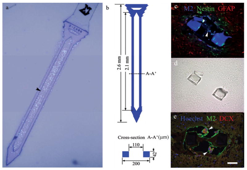
Figure 3.

A neural stem cell-seeded probe (a), and associated dimensions (b). Cells are Hoechst-stained and nuclei appear fluorescent blue (a). Cross-section views of neural stem cell-seeded probes 1 day following implantation from two animals (c-e). The companion brightfield DIC image for (c) is shown in (d) for reference due to autofluorescence of the parylene probe. Graft cells (M2-labeled) were associated with nestin (c), GFAP (c), and doublecortin (DCX, e) expression. Alginate also autofluoresces green. Scale = 50 microns for (c)-(e).
