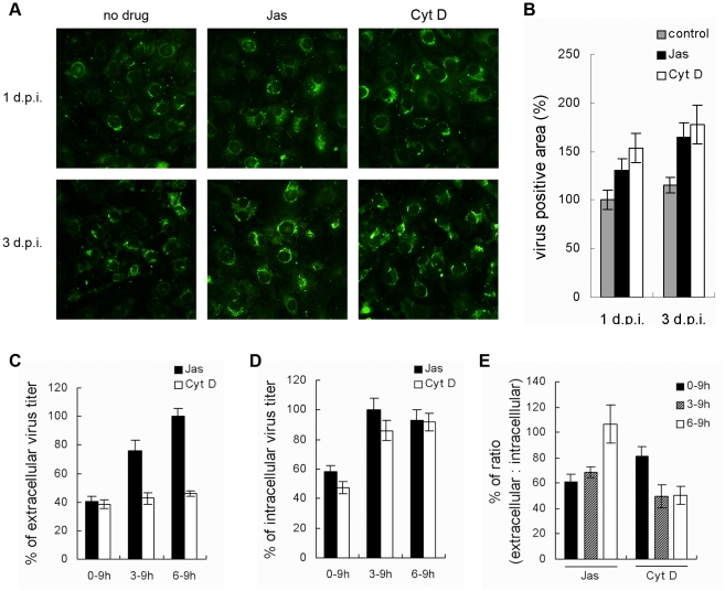
Figure 3. Actin is required for DV2 production and release.
(A, B) Accumulation of DV2 antigens in ECV304 cells continuously treated with actin inhibitors. ECV304 cells were infected with DV2 at 37°C for 1 h. Then cells were incubated with DMEM containing Cyt D (2 µM), Jas (100 nM), or DMSO and fixed at indicated time points. The cells were labeled with mouse anti-DV2 PAb, followed by FITC-conjugated anti-mouse IgG (A). (×400) Then the relative virus positive area was measured and the area of control cells was considered as 100% (B). (C, D, E) Time course of actin inhibitors effects on DV2 infection. Medium containing 0.1% DMSO, 100 nM Jas or 2 µM Cyt D was added to the ECV304 cells at the indicated time points post infection. The supernatant and cell samples were collected at 9 h p. i., and viral titers were measured by plaque assay (C and D). The numbers under each bar represent the treatment time of Jas or Cyt D. The ratio of extra- to intracellular viral titer is shown in percents (E). Experiments were performed in duplicate for at least three independent experiments.

