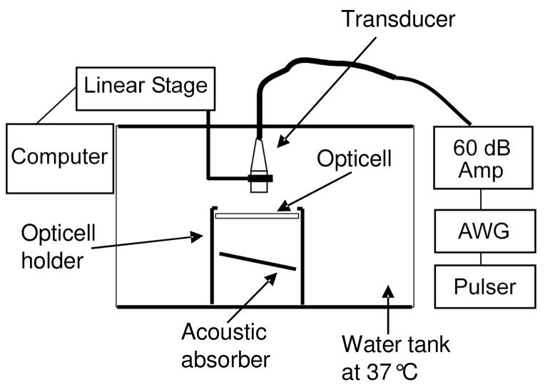
Figure 2.
In vitro experiments were performed in a water tank at 37°C. The transducer was oriented directly above the cells and its position was controlled with a linear motion controller. An acoustic neoprene absorber (7mm thick) below the Opticell was angled at 30 degrees to prevent standing wave formation. The PRF and pulse duration were set using a pulser and arbitrary waveform generator respectively. All pulses were amplified with a 60dB amplifier before reaching the transducer.

