Figure 3.
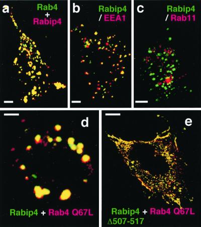
Rabip4 is localized in early sorting endosomes and gives enlarged vesicles with active Rab4. CHO cells were transiently transfected with pcDNA3-Rabip4 and pEGFP-Rab4 (a), pEGFP-Rabip4 (b and c), pEGFP-Rabip4 and pcDNA3 myc-Rab4 Q67L (d), or pEGFP-Rabip4 Δ(507–517) and pcDNA3 myc-Rab4 Q67L (e). Rabip4 is detected by using anti-Rabip4 antiserum and Texas Red-coupled anti-rabbit Ig (a) and myc-Rab4 Q67L is detected with mAb anti-myc followed by Texas Red-coupled anti-mouse Ig (d and e). Cells overexpressing GFP-Rabip4 were incubated with mAb anti-EEA1 (b) or with anti-Rab11 purified polyclonal Ig (c), followed by Texas Red-coupled anti-species Ig. Rab11 labeling was visible only in sections corresponding to the top of the cells (c), whereas no labeling was obtained when nonimmune Ig was used. The figures show merged images of green (GFP-labeled proteins) and red labeling obtained for the same section of representative CHO cells, with yellow color resulting from the overlay of green and red. (Bars = 1 μm.)

