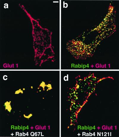
Figure 5.
Glut 1-myc distribution in cells expressing GFP-Rabip4. WT (a) or stably expressing GFP-Rabip4 (b–d) CHO cells were transiently transfected with pCis2 Glut 1-myc alone (a and b) or together with pCis2 Rab4 Q67L (c) or pCis2 Rab4 N121I (d). Three days after transfection, cells were fixed and permeabilized. Glut 1-myc is detected with anti-myc mAb and Texas red-coupled anti-mouse Ig. The merged images corresponding to green Rabip4 and red Glut 1-myc of the same confocal section are shown. Cells overexpressing Rab4 were identified by using anti-Rab4 serum and Cy5-coupled anti-rabbit Ig (data not shown). (Bar = 1 μm.)

