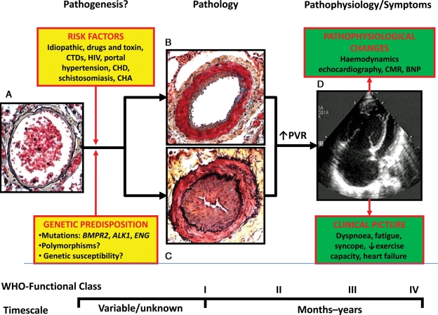
Figure 1.
Schematic representation of pathogenesis, pathology, pathophysiology, and symptoms in pulmonary arterial hypertension. (A) Normal small distal pulmonary artery: the thin wall is constituted by a single elastic lamina and a thin layer of smooth muscle cells; a large lumen with red blood cells is also shown. (B) Distal pulmonary artery in pulmonary arterial hypertension: increased thickness of the media due to hypertrophy and hyperplasia of smooth muscle cells and moderate lumen reduction are present. This picture may represent an initial phase of the disease and/or the prevalent changes in patients responding to vasoreactivity tests. (C) Distal pulmonary artery in pulmonary arterial hypertension: increased thickness of the media and also of the intima due to proliferation/migration of myofibroblasts and fibrosis are present. Severe lumen reduction is also shown. This picture may represent an advanced phase of the disease and/or the prevalent changes in patients not responding to vasoreactivity tests. (D) Echocardiographic four-chamber view in pulmonary arterial hypertension: Severe dilatation of the right atrium and ventricle and reduction in size of the left ventricle are shown. ALK1, activin-like kinase-type 1 gene; BNP, brain natriuretic peptide; BMPR2, bone morphogenetic protein receptor type 2 gene; CHA, chronic haemolytic anaemia; CHD, congenital heart disease; CTD, connective tissue diseases; ENG, endoglin gene; HIV, human immunodeficiency virus; CMR, cardiac magnetic resonance; PAH, pulmonary arterial hypertension; PVR, pulmonary vascular resistance; WHO, world health organization. The pathological pictures are a courtesy of Dr Carol Farver, Cleveland Clinic.

