Figure 3.
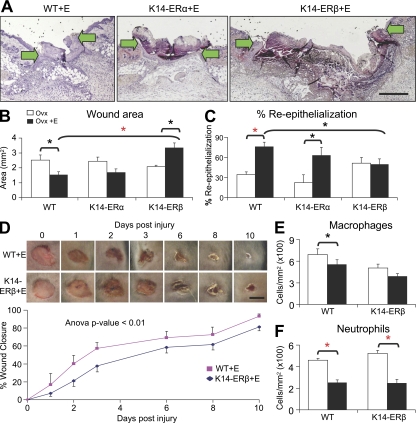
Epidermal-specific ERβ null (K14-ERβ) mice phenocopy ERβ−/− mice. (A) Representative H&E-stained sections from estrogen (E)-treated wild-type (WT), K14-ERα, and K14-ERβ mice. (B and C) Estrogen treatment of K14-ERβ mice specifically delays healing, shown by increased wound area (B) with no increase in re-epithelialization (C). (D) Temporal monitoring of excisional wound closure reveals that delayed healing (K14-ERβ) is maintained through later time points. (E and F) Quantification of wound inflammatory cell numbers reveals antiinflammatory effects for estrogen on neutrophils in WT and K14-ERβ (E). Data shown as mean ± SEM are representative of two independent experiments (n = 6). (A) “K14-ERβ+E” image has been auto stitched from two individual images. Bars: (A) 400 µm; (D) 4 mm. Black asterisk, P < 0.05; red asterisk, P < 0.01.

