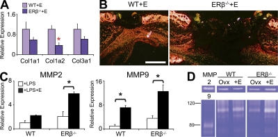
Figure 6.
Altered matrix deposition and protease activity in ERβ null (ERβ−/−) mice. (A and B) Wound collagen content determined by expression of major collagen species (qPCR) and Picro-Sirius staining was reduced in estrogen (E)-treated ERβ−/− mice. (C) E-treated peritoneal macrophages from ERβ−/− mice display increased MMP-2 and MMP-9 expression. (D) Via zymography, MMP-9 activity was strongly increased in E-treated ERβ−/− wounds. Data from two independent experiments are shown as mean ± SEM using cells in triplicate wells (C) or four animals per group (A). (B and D) Representative of at least four animals per group from three independent experiments. (B) “ERβ−/−+E” image has been auto stitched from two individual images. Bar, 800 µm. Black asterisk, P < 0.05; red asterisk, P < 0.01.

