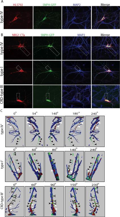
Fig 5. Subcellular localization of NRG1 type IV in hippocampal neurons.
A) Dissociated hippocampal neurons co-transfected with NRG1 type IV and SNPH-GFP, and live labeled with type IV antibodies. Cells were also stained for the dendritic marker MAP2 after permeabilization. Type IV immunolabeling (red) colocalizes with the dendritic marker MAP2 (blue) and is strongest in the proximal region of dendrites. (B) Hippocampal neurons were co-transfected with NRG1 type I, type IV or type III and SNPH-GFP (green), permeabilized, and incubated with antibodies against NRG1 (red) and MAP2 (blue). Type I and IV immunoreactivity is restricted to neuronal somas and dendrites, and absent from SNPH positive axons. In contrast, NRG1 CRD-type III is localized to the cell somata and dendrites but it also was targeted to the axon (green). (C) Three dimension reconstruction of the area obtained by the dash box in B. Axons (green) are indicated by black arrows. Type IV and I immunolabeling can be observed in the proximal region of the axon (white arrowheads) but not in more distal region. Unlike type IV and I, CRD-type III is expressed in the axon (white filled arrowheads).

