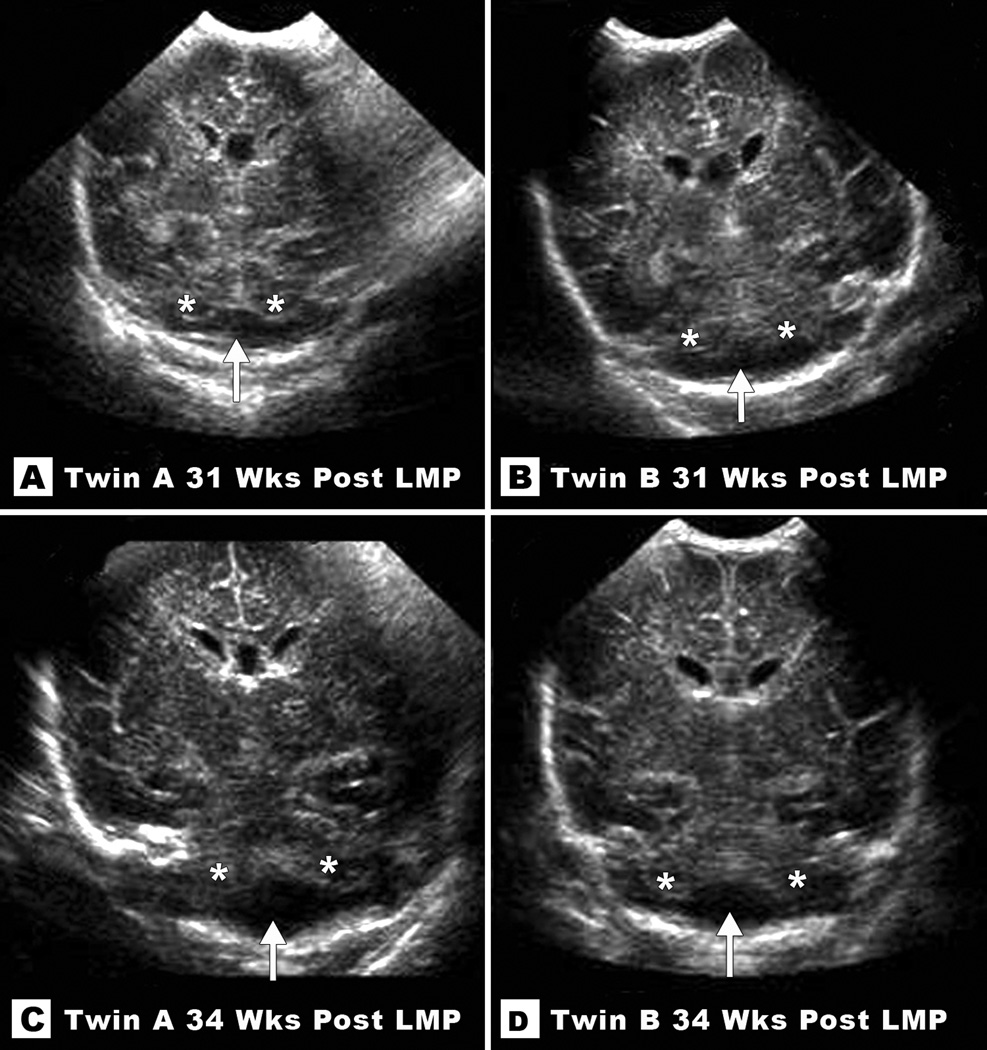
Figure 2.
Coronal sections from neonatal ultrasounds at 31 weeks (top) and 34 weeks (bottom) post-LMP in Twin A (left) and Twin B (right) reveal subtle cerebellar hypoplasia. Note empty cistern (marked with an arrow) in the lower half of the posterior fossa due to absent cerebellar tissue (remaining cerebellar hemispheres marked with asterisks).

