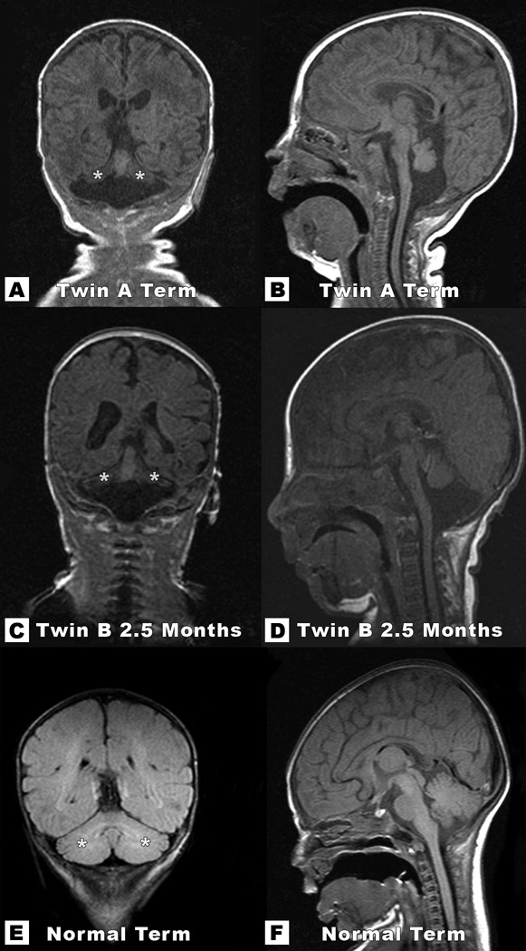
Figure 3.

Coronal T1-weighted images of Twin A at 2.5 months of life (A and B) and Twin B at 5 months, 2.5 months corrected for prematurity (C and D) depict severe hypoplasia of both cerebellar hemispheres. Note the typical “winging” appearance of cerebellar hemispheres (marked with asterisks), which are compared with similar images in a term neonate (E and F). Mid sagittal T1-weighted images of each twin (Figures B and D) show significant volume loss of the pons and relative preservation of the vermis, with much better preservation of the cerebellar vermis than the cerebellar hemispheres.
