
Figure 1.
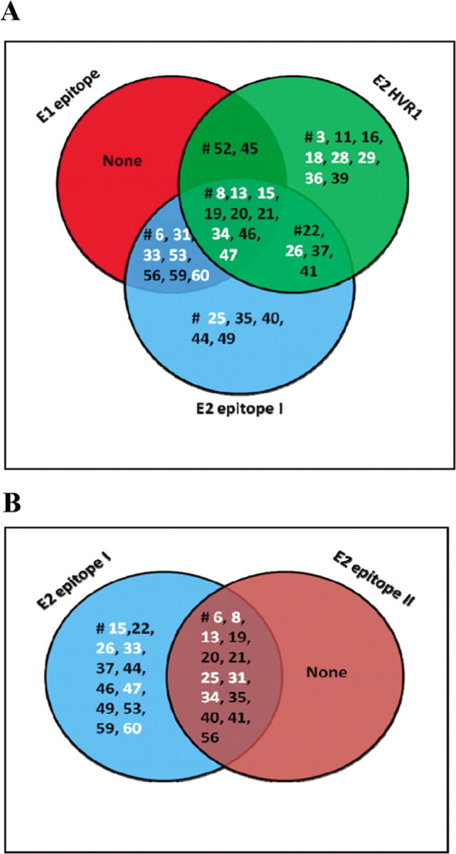
Venn diagrams representing unique and shared epitope recognition of hepatitis C virus envelope glycoproteins E1 and E2 by serum samples from volunteers who were vaccinated with E1/E2. The reactivity level of a panel of serum samples from vaccinees was tested at a 1: 100 dilution against biotinylated peptides representing linear epitopes of E1 or E2 immobilized on an avidin-coated enzyme-linked immunosorbent assay (ELISA) plate. The E1 epitope encompassing amino acid residues 313–327 (Ile-Thr-Gly-His-Arg-Met-Ala-Trp-Asp-Met-Met-Met-Asn-Trp-Ser-OH), the E2 hypervariable region 1 (HVR1) epitope encompassing amino acid residues 384–411 (Glu-Thr-His-Val-Thr-Gly-Gly-Ser-Ala-Gly-His-Thr-Val-Ser-Gly-Phe-Val-Ser-Leu-Leu-Ala-Pro-Gly-Ala-Lys-Gln-Asn-OH), the E2 epitope 1 encompassing amino acid residues 412–419 (Gln-Leu-Ile-Asn-Thr-Asn-Gly-Ser-Trp-His-Ile-Asn-Ser-Thr-Ala-OH), and the E2 epitope 2 encompassing amino acid residues 434–446 (Leu-Asn-Thr-Gly-Trp-Leu-Ala-Gly-Leu-Phe-Tyr-Gln-His-Lys-Phe-OH) were immobilized (20 ng per well) on avidin-coated ELISA plates. The plates were incubated with 1:50 dilutions of test serum samples overnight at 4°C and washed, and bound antibody was detected by adding streptavidin-horseradish peroxidase- conjugated antibody to human immunoglobulin and peroxidase substrate. The color intensity was measured by absorbance at 492 nm. Results that displayed reactivity with an increase in optical density of >2-fold compared with a matched preimmune or placebo control serum sample were considered positive. The overlapping regions represent serum samples that concomitantly recognized epitopes on E1 and E2 (A). Serum samples that recognized E2-neutralizing epitope 1 and nonneutralizing epitope 2 are also shown (B). Serum samples that displayed neutralizing activity are labeled in white.