
Figure 6.
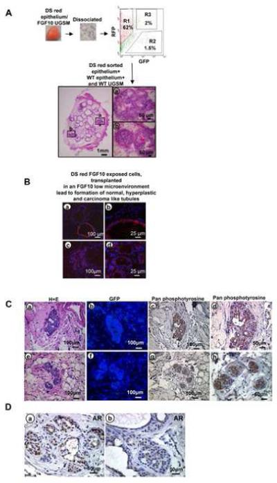
Serial transplantation of FGF10 induced adenocarcinoma
A: Schematic representation of transplantation experiments and histology of regenerated tubules.
B: Immunofluorescent analysis of regenerated tissue revealed formation of many WT and some red epithelial glands with a single cell layer resembling normal tubules (a&b), and multiple small red tubules adjacent to normal appearing WT glands reminiscent of well differentiated adenocarcinoma (c&d).
C: Histologic evaluation of transplanted tissue specimens revealed evidence of adenocarcinoma (a&e) and absence of contaminating FGF10 expressing stromal cells demonstrated by lack of GFP staining (b&f). The cancerous regenerated tissue revealed strong phosphotyrosine activation after withdrawal of retrovirally infected FGF10 UGSM cells (c&d, g&h).
D: Persistent strong nuclear localization of AR was observed in cancerous areas of the transplanted regenerated grafts (a&b).