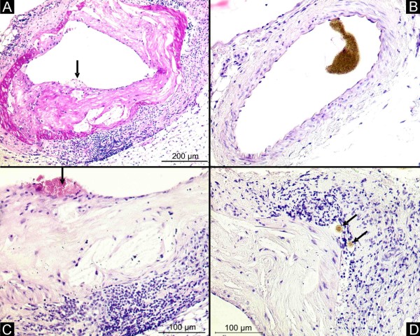
Figure 1.
Renal artery. Histology demonstrates concentric atherosclerotic plaques in the renal artery of the apoE-/-/LDL-/- double knockout mouse at the age of 80 weeks (A, magnification × 50). Control animals showed no atherosclerotic lesions in the renal artery (B, magnification × 50). In high-power fields, plaque rupture (C, black arrow, magnification × 100) and contrast enhanced Vasa vasorum (D, black arrow, magnification × 100) in the adventitia with large clusters of inflammatory cells occur.

