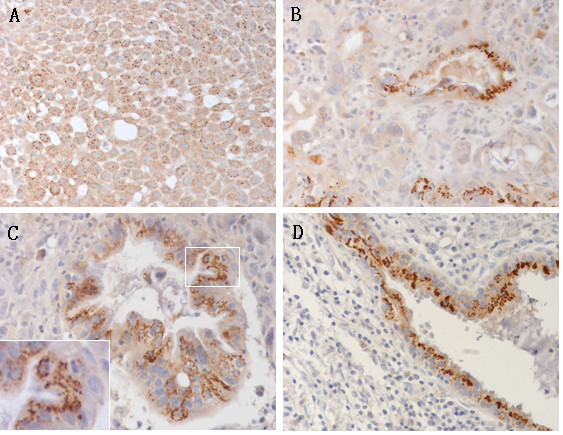
Figure 4.

Immunohistochemistry of Sp17 in cervical cancer tissues (A-C) and hyperplastic gland (D). The staining pattern of Sp17 was granulo in all the original cervical tissues and located in the cavosurface of adenocancer and the hyperplastic gland. A. squamous cancer; B, C, adenocancer; D. hyperplastic glands in the periphery of the lesions. Sp17 was localized in the cytoplasm but had different intensity. (A, B, D, 20×; C, 40× original magnification, 100× inset).
