
Figure 1.
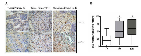
Immunologic detection of NF-κB p65 in SCCHN tissues. (a) Representative images of p65 immunostaining of a primary tumor with lymph node involvement (N+) (Tm), the corresponding lymph node contain metastases (Ln) from the same patient and a primary tumor without lymph node involvement (N-) (Tn). (b) Graphical representation of the percentage of p65-positive nuclei in the SCCHN tissue. The asterisk (*) denotes p < 0.01.