
Figure 2.
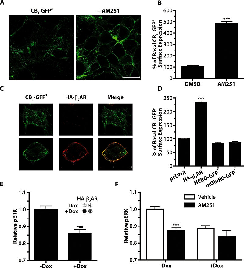
Co-expression of HA-β2AR reduces the constitutive activity of CB1-GFP2 in 293H cells. (A) Confocal images of 293H cells stably expressing CB1-GFP2 treated for 24 h with 0.05% DMSO vehicle (left panel) or 10 µM AM251 (right panel). Scale bar is 20 µm. (B) On-Cell Western quantitative measure of CB1 cell surface expression following 24 h AM251 treatment (10 µM) in 293H cells stably expressing CB1-GFP2. ***P < 0.001 compared with DMSO vehicle; n= 4–6. (C) Confocal images of 293H cells transfected with CB1-GFP2 and HA-β2AR. Left panels show GFP2 fluorescence, middle panels are anti-HA immunofluorescence utilizing a Cy3-conjugated secondary antibody, and the right panels are the merged images. Scale bar is 20 µm. (D) On-Cell Western quantitative measure of CB1-GFP2 cell surface expression in 293H cells stably expressing CB1-GFP2 and transiently transfected with pcDNA, HA-β2AR, HERG-GFP2 or mGluR6-GFP2. ***P < 0.001 compared with pcDNA transfected cells; n= 4–18. (E) Basal pERK levels in CB1-GFP2/TreHA-β2AR cells without and with Dox pretreatment (10 µg·mL−1, 24 h) to induce expression of HA-β2AR. ***P < 0.001; n= 20. Inset shows On-Cell Western using an anti-HA primary antibody to measure HA-β2 expression without or with Dox. (F) pERK levels in CB1-GFP2/TreHA-β2AR cells treated with 0.05% DMSO vehicle (open bars) or AM251 (1 µM, 10 min, solid bars) in cells without or with Dox pretreatment (10 µg·mL−1, 24 h). ***P < 0.001 compared with respective vehicle controls; n= 20–34. AM251, N-(piperidin-1-yl)-5-(4-iodophenyl)-1-(2,4-dichlorophenyl)-4-methyl-1H-pyrazole-3-carboxamide; DMSO, dimethylsulphoxide; Dox, doxycycline; ERK, extracellular signal-regulated kinase; HERG, human ether-a-go-gorelated gene; mGluR6, metabotropic glutamate receptor 6.