
Figure 3.
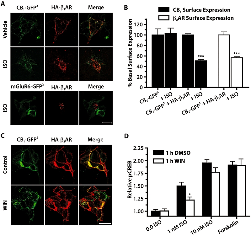
HA-β2AR and CB1-GFP2 are co-internalized when exposed to either isoprenaline (ISO) or WIN. (A) Confocal images of 293H cells transiently transfected with CB1-GFP2 or mGluR6-GFP2 and HA-β2AR treated for 30 min with vehicle or 10 µM isoprenaline. Left panels show GFP2 fluorescence, middle panels are anti-HA immunofluorescence utilizing a Cy3-conjugated secondary antibody, and right panels are merged images. Scale bar is 20 µm. (B) On-Cell Western quantitative measure of CB1-GFP2 or HA-β2AR cell surface expression in 293H cells stably expressing CB1-GFP2 and transiently transfected with either pcDNA (CB1-GFP2 bars) or HA-β2AR. Cells were treated with either H2O vehicle (labelled CB1-GFP2 + HA-β2) or isoprenaline (10 µM) for 30 min. ***P < 0.001 compared with CB1-GFP2 + HA-β2AR vehicle-treated groups; n= 6. (C) Confocal images of HEK 293H cells transiently transfected with CB1-GFP2 and HA-β2AR, upper panels are untreated controls, and lower panels are treated with WIN (10 µM, 30 min). Scale bar is 20 µm. (D) Relative pCREB levels in CB1-GFP2/TreHA-β2AR cells pretreated with Dox (10 µM, 24 h), treated for 1 h with either DMSO (0.05%) or WIN (10 µM), followed by 30 min treatment with isoprenaline (0–10 nM), or forskolin (10 µM). *P < 0.05 compared with respective 1 h DMSO treatments; n= 12–20. CREB, cyclic AMP response element binding protein; DMSO, dimethylsulphoxide; Dox, doxycycline; HEK, human embryonic kidney; mGluR6, metabotropic glutamate receptor 6; WIN, WIN 55,212-2.